


Internet-Enabled Robotic Microscope
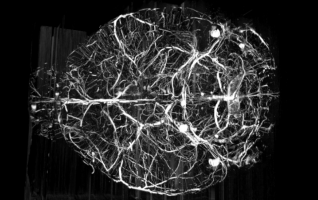
Blood vessel from a mouse brain cortex.
Cerebellar Purkinje cells
Blood Vessel 3D Volume Visualization
3D Printed Mouse Brain Blood Vessel
Brain Research through Digitizing Brain Samples
The Brain-Inspired Intelligent Systems (BI2S) Laboratory is part of the Department of Electrical and Computer Engineering at Kettering University. The BI2S Lab houses the Internet-Enabled RObotic Microscope (IEROM) powered by Knife-Edge Scanning Microscope (KESM) technology. The IEROM is a robotized sectioning and sub-micron imaging instrument for investigating biological tissue volumes.
Instrumentation
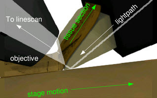
Knife-Edge Scanning Microscopy not only preserves image registration throughout the depth of the specimen block but also isolates the tissue above the knife from that below to eliminate undesirable events.
Teravoxel Image Processing
Teravoxel-sized 3D image stacks become common as sectioning microscopy technologies advances. Due to the immense size of the data sets, fast, accurate, and automated image processing pipelines are essential for further quantitative analysis of the data.
Modeling
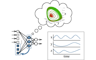
Study of how internal brain properties invisible to evolution can still evolve as environment changes.